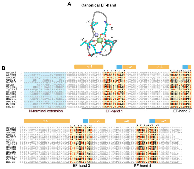
Figure 1.
Overview of the EF-hand Ca2+ binding domains in different centrins. (A) Ca2+ coordination by the canonical EF-hand (PDB: 1CLL). The Ca2+ ion is coordinated in a pentagonal bipyramidal configuration by ligands indicated by their position in the coordination geometry (X, Y, Z, −X, −Y and −Z). NH groups of coordinating amino acids are indicated in dark blue, oxygen atoms in red, the Ca2+ ion in green and the coordinating water molecule in violet. (B) Protein sequence alignment of centrins from different organisms. The N-terminal extension (light blue box) and the central 12 residues in the EF-hand domains (orange boxes) are highlighted. Within the EF-hands, Ca2+ chelating residues are represented in orange while the other most common residues are represented in black. Secondary structural elements derived from the 3D structure of human CaM (PDB: 1CLL), α-helices (orange) and β-sheets (light blue) are displayed on the top of the alignment.

