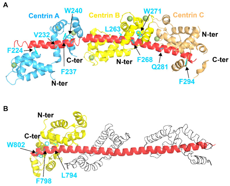
Figure 3.
Crystal structures of (A) the complex between SFI1 and CDC31 molecules and (B) the complex between SAC3, SUS1, and CDC31. (A) Crystal structure of three yeast centrins CDC31 (light blue, yellow and orange) bound to SFI1 (PDB: 2DOQ). Ca2+ ions are indicated by smudge green spheres. The anchoring residues for the interaction of SFI1 with CDC31 are indicated in cyan. (B) 3D structure of the complex CDC31 (yellow), SAC3 (red) and SUS1 (transparent) (PDB: 3FWC). The key residues constituting the large hydrophobic surface for the interaction of SAC3 with CDC31 are indicated in cyan.

