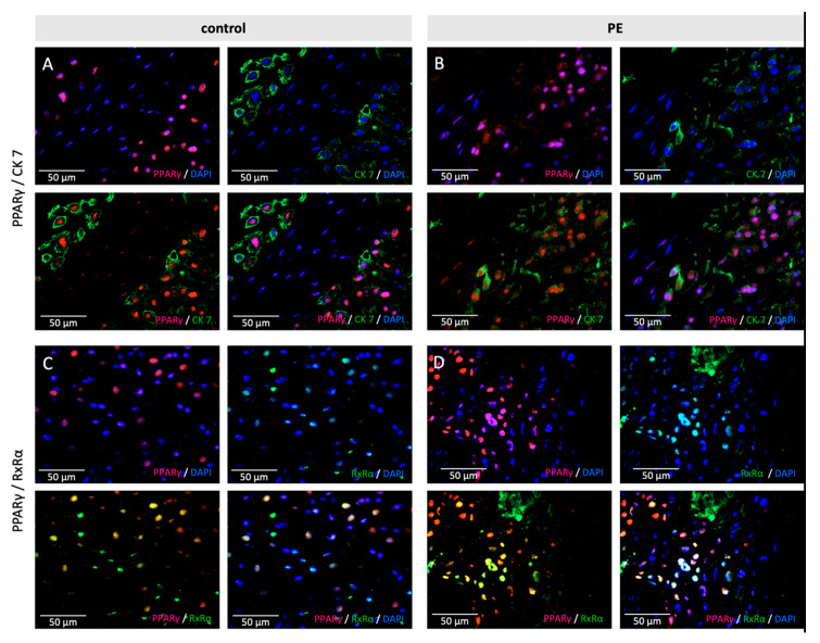
Figure 3.
Examples of staining results of the immunofluorescence of PPARγ with CK7 (A,B) and RxRα (C,D), in control (A,C) and PE (B,D) placentas. Single immunofluorescence staining of PPARγ (pink). Single immunofluorescence staining CK7 or RxRα (green). Double immunofluorescence staining of PPARγ (A/B) and H3K4me3 (C/D) (red) and PPARγ (green). DAPI as nucleus staining (blue). Scale bar 100 µm.

