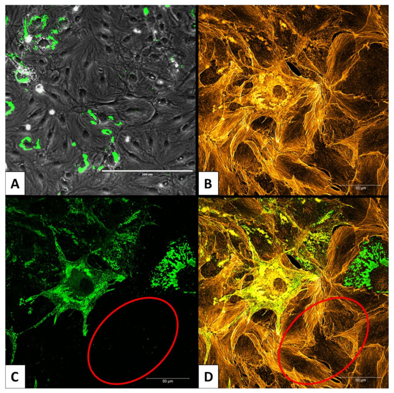
Figure 4.
Persistently infected MoLu Prim_EBOV cells. Overview of persistently infected MoLu Prim_EBOV cells 143 dpi; scale bar: 200 µm (A). Enlargement of MoLu Prim_EBOV cells; scale bar: 50 µm. Stained actin filaments (B); stained EBOV-NP (C). Overlay B and C (D). Area with uninfected cells (red ellipse), EBOV-NP (green), actin filaments (orange).

