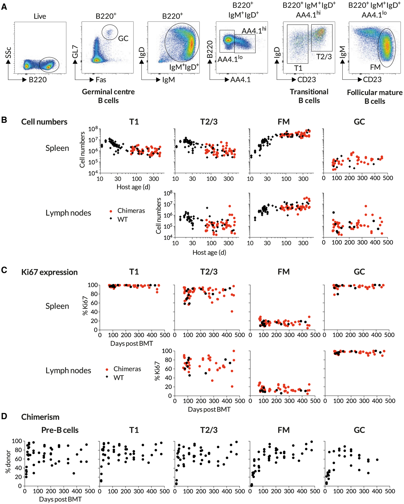
Figure 1. Busulfan Chimeric Mice Exhibit Normal Peripheral B Cell Compartments.

Busulfan chimeras were generated as described in STAR Methods (n = 47) and compared with WT controls (n = 74). Data are pooled from multiple experiments.
(A) Gating strategy to identify transitional, follicular mature, and germinal center B cells.
(B) Comparing the sizes of B cell subsets in WT control mice and busulfan chimeras.
(C) Comparing proliferative activity in WT and busulfan chimeric mice, using Ki67 expression.
(D) Host-derived B cells are gradually replaced by donor-derived cells over time. Scatter derives largely from variation in levels of stable bone marrow chimerism achieved in treated mice.
See also Figure S1.
