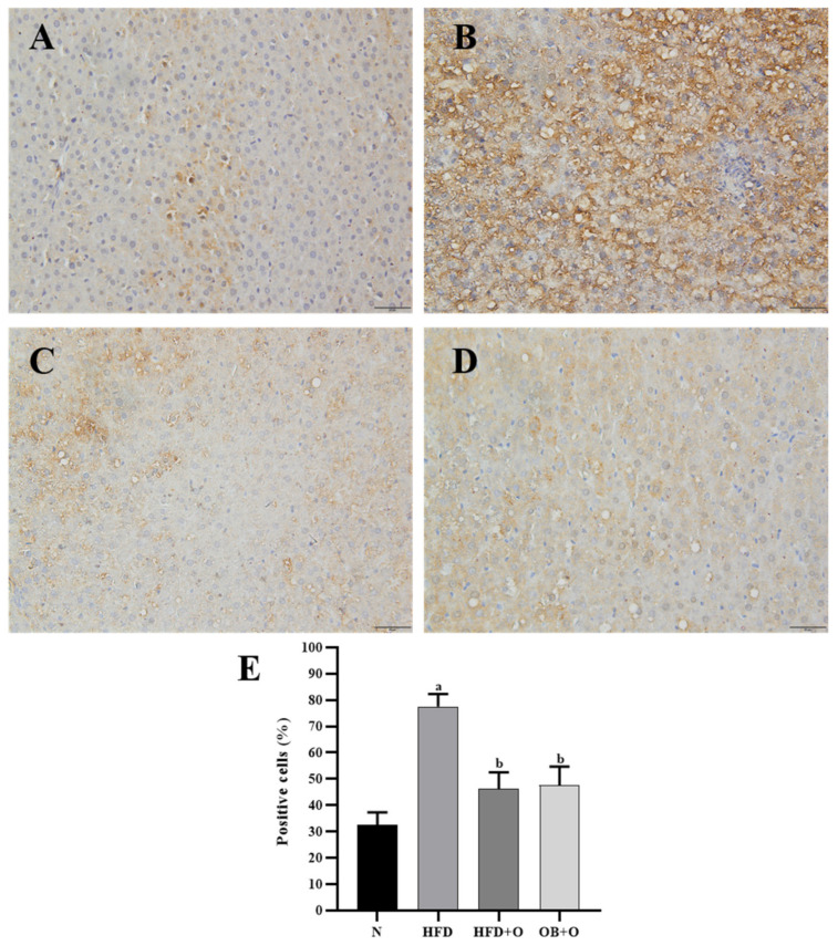
Figure 6.
(A–E). Immunohistochemical staining of Keap1 expression in liver sections. N, normal control; HFD, high-fat diet; HFD + O, high-fat diet + orlistat 10 mg/kg/day (protective model); OB + O, obese+orlistat 10 mg/kg/day (therapeutic model); Keap1, Kelch-like ECH-associated protein 1. (A) The N group shows less accumulation of Keap1 in the cytoplasm. (B) More Keap1 was concentrated in the cytoplasm of the HFD group than in the N group. (C,D) This concentration morphology was reduced after orlistat administration in the HFD + O and OB + O groups. Magnification, ×400. (E) Quantification of Keap1-positive cells (%). Data are expressed as mean ± SEM, n = 6/group. One-way ANOVA, followed by Tukey post-hoc test. a p < 0.05 vs. N group, b p < 0.05 vs. HFD group.

