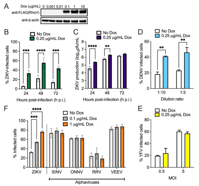
Figure 3.
RhoV enhances ZIKV and DENV but not alphavirus infection in SNB-19 cells. (A) Inducible RhoV SNB-19 cells were treated with different amounts of Dox (0, 0.001, 0.01, 0.1, 1, and 10 µg/mL) for 24 h to induce the expression of N-terminally 3xFLAG-tagged RhoV through the ePiggyBac transposon system. RhoV and β-actin (loading control) protein expression determined by immunoblotting with FLAG and β-actin antibodies. The data are representative of two independent experiments. (B) Inducible RhoV SNB-19 cells were treated with or without 0.25 µg/mL Dox for 24 h prior to 1 h adsorption with ZIKV (MOI = 0.5 PFU/cell) and harvested at 24, 48, and 72 h.p.i. to quantify infection levels. Dox was added back to the media during the course of infection. ZIKV infected cells were then fixed and permeabilized to stain with the pan-flavivirus envelope antibody prior to flow cytometry analysis. The data are combined from two independent experiments. (C) Supernatant of infected cells from (B) was collected at the same timepoints and titered on Vero cells by plaque assay. The data are representative of two independent experiments. Inducible RhoV SNB-19 cells were treated with or without 0.25 µg/mL Dox for 24 h prior to 1 h adsorption with (D) DENV-GFP (1:10 or 1:5 dilution) or (E) YFV 17D Venus (MOI = 0.5 or 5 PFU/cell) and harvested at (D) 72 h.p.i. or (E) 48 h.p.i. to quantify infection levels. Dox was added back to the media during the course of infection. YFV- and DENV-infected cells were fixed prior to flow cytometry analysis for GFP/Venus expression. The data are representative of two independent experiments. (F) Inducible RhoV SNB-19 cells were treated with 0.1 and 1 µg/mL Dox 24 h prior to 1 h adsorption with ZIKV (MOI = 0.5 PFU/cell), and GFP-expressing SINV (MOI = 1 PFU/cell), ONNV (MOI = 1 PFU/cell), VEEV (MOI = 0.5 PFU/cell), and RRV (MOI = 5 PFU/cell) and harvested at 24 h.p.i. to quantify infection levels. Dox was added back to the media during the course of infection. ZIKV infected cells were then fixed and permeabilized to stain with the pan-flavivirus envelope antibody as above, while alphavirus-infected cells were fixed prior to flow cytometry analysis for GFP expression. The data are combined from two independent experiments. Asterisks indicate statistically significant differences (two-way ANOVA and (B–E) Sidak’s or (F) Dunnett’s multiple comparisons test: **, p < 0.01; ***, p < 0.001; ****, p < 0.0001).

