Figure 5.
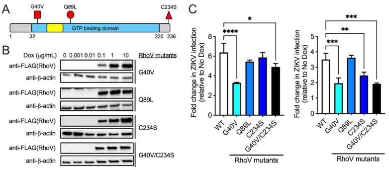
RhoV GTPase domain contributes to its proviral effects during ZIKV infection. (A) Schematic of RhoV protein domains and mutations introduced (red flags). The GTP binding/hydrolysis domain (blue), effector or switch I domain (yellow), and N- and C-terminal extension regions (gray) are shown. Adapted from [18]. (B) SNB-19 cells were treated with different amounts of Dox (0, 0.001, 0.01, 0.1, 1 and 10 µg/mL) for 24 h to induce the expression through the ePiggyBac transposon system of N-terminally 3×FLAG-tagged RhoV WT or mutants that are dominant-activated, GTPase-defective (G40V, Q89L), mislocalized (C234S), or both (G40V/C234S). Mutant RhoV and β-actin (loading control) protein expression was determined by immunoblotting with FLAG and β-actin antibodies. The data are representative of two independent experiments. (C) Two independent experiments are shown here (left and right). Inducible RhoV (WT or mutants) SNB-19 cells were treated with or without 0.25 µg/mL Dox for 24 h prior to 1 h adsorption with ZIKV (MOI = 0.5 PFU/cell) and harvested at 24 h.p.i. to quantify infection levels. Dox was added back to the media during the course of infection. ZIKV infected cells were then fixed and permeabilized to stain with the pan-flavivirus envelope antibody prior to flow cytometry analysis. Infection levels of RhoV WT and mutant cell lines were normalized to that of the respective untreated condition (no Dox) and reported as fold changes. Asterisks indicate statistically significant differences (one-way ANOVA and Dunnett’s multiple comparisons test: *, p < 0.05; **, p < 0.01; ***, p < 0.001; ****, p < 0.0001).

