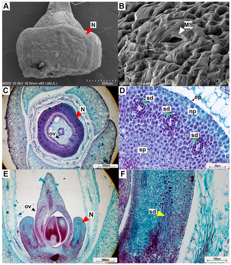
Figure 6.
Photographs from micromorphological investigation of the floral nectary of Argyreia siamensis: (A) the entire floral nectary (which surrounds the ovary) and (B) surface of the floral nectary viewed under a scanning electron microscope; (C) transverse section of a mature flower showing the nectary and (D) transverse section showing close-up details of the nectary tissue; (E) longitudinal section of the flower and (F) longitudinal section of the floral nectary showing a close-up view of the secretory duct. In photos C-F, sections were stained with safranin-O and fast green. Abbreviations: N = nectary, MS = modified-stomata, ep = epidermis, ov = ovary, np = nectariferous parenchyma, sp = subnectariferous parenchyma, sd = secretory duct. Scale bars: (A) 500 µm; (B) 40 µm; (C) 500 µm; (D) 50 µm; (E) 500 µm; (F) 100 µm.

