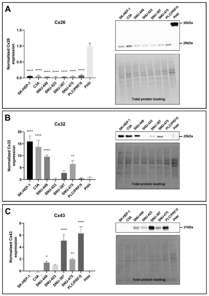
Figure 3.
Cx26 (A), Cx32 (B) and Cx43 (C) protein expression in liver cancer cell lines and primary human hepatocytes (PHH). Cancer cell lines (n = 1, N = 3) were grown to 100% confluence, while PHH were used in suspension for protein extraction. Immunoblotting and visualization were done with the Pierce™ ECL Western Blotting Substrate kit (Thermo Fisher Scientific, Waltham, MA, USA) on a ChemiDocTM MP imaging system. All signals were divided by their respective total protein loading signal and normalized by the sum of all data points in a replicate [42]. Unlike Cx43, which was not expressed by PHH, Cx26 and Cx32 are expressed relative to their expression in PHH. Data are expressed as mean ± standard deviation with * p ≤ 0.05, ** p ≤ 0.01 and **** p ≤ 0.0001 compared to the PHH control.

