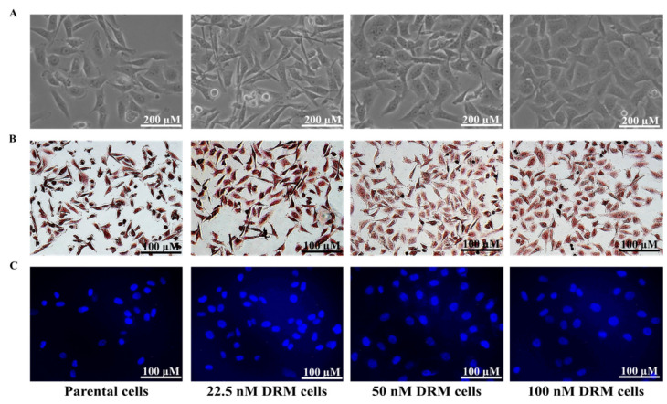
Figure 2.
Morphological analysis of DRM cells (A) Morphological analysis performed under a light microscope 48 h after cells were seeded. DRM cells showed an increase in cell size and cell-to-cell communication. The cells changed from spindle-like structures to cobblestone-like giant cells as acquiring resistance to doxorubicin (Magnification, ×200; scale bar, 200 µm) (B) Whole-cell morphology analyzed with Mayer’s staining. DRM cells showed morphological changes with acquiring resistance to higher concentrations of doxorubicin. (Magnification, ×100; scale bar, 100 µm) (C) DAPI staining showing the nuclear morphology of DRM cells. DRM cells showed an increase in nuclear size with advancement in doxorubicin resistance. Results were confirmed by three independent experiments. (Magnification, ×100; scale bar, 100 µm). DRM: Doxorubicin-resistant MDA-MB-231.

