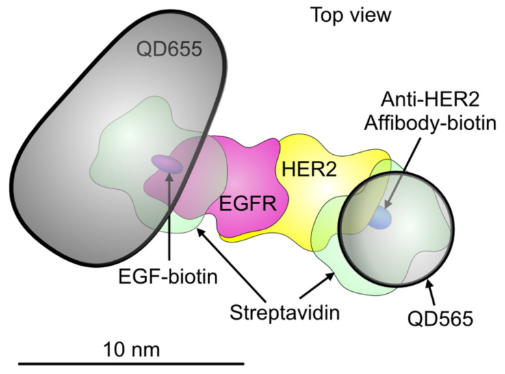
Figure 1.
Schematic model depicting an EGFR-HER2 heterodimer with two bound quantum dots (QDs). A QD655 (the bullet-shaped core is shown) is bound to EGFR via streptavidin and an EGF-biotin linker. A smaller QD565 (spherical shape) is bound to HER2 through a streptavidin and an Affibody-biotin compound. The published molecular model of the EGFR-HER2 heterodimer was used [45]. The other used molecular structures (5WB7, 3MZW, and 1STP) were obtained from the Protein Data Base. Dimensions are drawn to scale.

