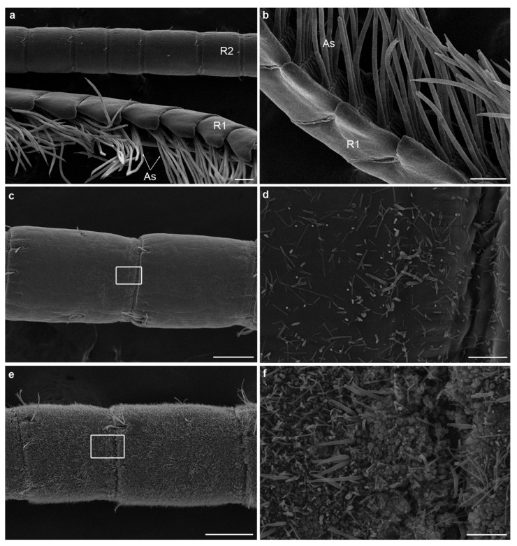
Figure 7.
Scanning electron micrographs of Palaemon serratus (juveniles) before (Ps1, Ps2) and after (Ps10, Ps11) grooming experiments. (a,c,e) Antennules of Ps10 (a), Ps1 (c) and Ps2 (e) specimens showing gradation from absent (a), to moderate (c) and intense (e) bacterial fouling on the antennal segments. (a) Antennules of Ps10 specimen, showing the two ramus and the aesthetascs completely devoid of bacterial fouling; (b) close-up on the aesthetascs of Ps11 specimen completely devoid of bacterial fouling; (c,d) antennae of Ps1 specimen with a light fouling of bacteria. Frame in (c) is enlarged in d showing the bacterial morphological variety, with thick and thin filamentous bacteria, some rods and cocci; (e,f) antennules of Ps2 specimen with very dense fouling covering the entire surface of the segments. Frame in (e) is enlarged in (f), showing bacterial density and morphological diversity. As: aesthetascs, R1 and R2: the two rami of the lateral antennular flagellum. Scale bars: (a–c,e) = 100 µm; (d,f) = 10 µm.

