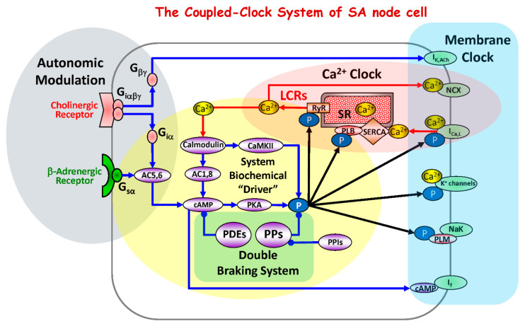
Figure 1.
Schematic diagram of the coupled-clock system that includes four major sub-systems or functional modules: the sarcoplasmic reticulum, the Ca2+ clock; sarcolemmal ion channels and transporters, the membrane clock; biochemical drivers; and the autonomic modulation of the system components. The Ca2+ clock cycles Ca2+ via SR Ca2+ pump (SERCA) and Ca2+ release channels (RyRs). The membrane clock generates APs and interacts with the Ca2+ clock via multiple Ca2+-dependent mechanism, including NCX current that accelerates the diastolic depolarization. The system’s biochemical driver is cAMP, which is generated by Ca2+-activated AC1and AC8 and leads to activation of PKA. PKA and CaMKII increase phosphorylation of clock proteins (black arrows). The autonomic nervous system modulates the clock system via G protein-coupled receptor signaling. The cAMP level is kept in check by PDEs. The focus of the present study is to determine whether PPs, by keeping clock protein phosphorylation levels in check, form the double braking system with PDEs. Abbreviations: IK,Ach, acetylcholine-activated K+ current; NCX, Na+/Ca2+ exchanger; ICaL, L-type Ca2+ current; K+ channels, potassium channels; If, hyperpolarization-activated current; Ca2+, calcium ions; LCRs, local submembrane Ca2+ releases; RyR, ryanodine receptors; SR, sarcoplasmic reticulum; SERCA, SR Ca2+ ATPase; CaMKII, calcium-calmodulin-dependent protein kinase II; PLM, phospholemman; PLB, phospholamban; P, phosphorylation; PKA, protein kinase A; cAMP, cyclic adenosine 3′,5′-monophosphate; AC, adenylyl cyclase; Ggsα, G-protein coupled receptors stimulatory alpha subunit; Giα, G-protein coupled receptors inhibitory alpha subunit; Giαβγ, G-protein coupled receptors inhibitory alpha, betta, gamma subunits; Gβγ, G-protein coupled receptors beta, gamma subunits; PPs, phosphoprotein phosphatases; PPIs, phosphoprotein phosphatase inhibitors; PDEs, phosphodiesterases.

