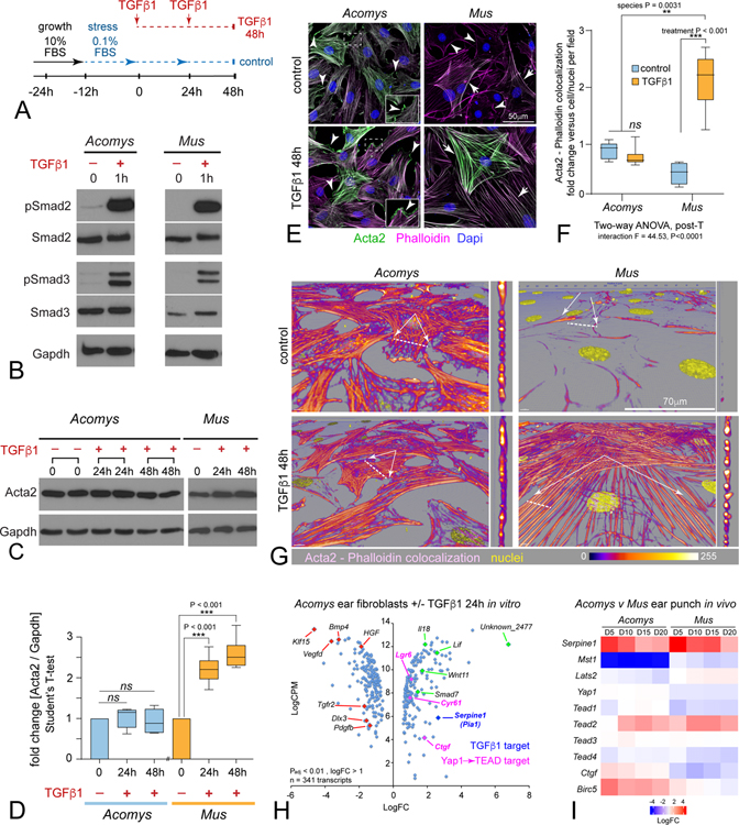
Figure 2: Acomys primary ear tissue fibroblasts respond to fibrokine signaling but resist terminal myofibroblast differentiation.
(A) Diagram for MF induction assay in response to TGFβ (2ng/ml).
(B) Western blot for Smad2 and Smad3 reveals phosphorylation in both Acomys and Mus primary ear fibroblasts after 1h TGFβ1.
(C) Western blot of Acta2 protein levels from Acomys and Mus in response to TGFβ1 at the indicated times: N ≥ 4 independent cultures, 2 replicates shown for Acomys.
(D) Quantification of Western blots reveals significant increase in Mus compared to no change in Acomys, two-way ANOVA, post-T as indicated; data are mean ± SD.
(E) Immunostaining Acta2 (green) in primary Acomys and Mus ear fibroblasts exposed to 48h TGFβ1 co-stained with Phalloidin (magenta), and Dapi (blue), even retaining Acta2+ lamellipodia with TGFβ1 exposure (insets), by contrast to Mus.
(F) Quantification of Acta2/Phalloidin colocalization levels after 48h TGFβ1 reveals similar significant increases in Mus compared to no change in Acomys, as indicated (see also Figure S3); data are mean ± SD.
(G) Confocal rotated higher power rendered views of individual fibroblasts reveal minimal structural or morphological differences in Acomys Acta2/Phalloidin colocalized short filaments by contrast to MFs with thick F-actin stress fibers spanning flattened cells in Mus.
(H) Scatter plot of RNAseq analysis (bullk) comparing Acomys DFs treated with TGFβ1+ versus stressed controls (24h): note lack of Acta2 transcriptional up- or down-regulation: Green, candidate up-regulated genes; Red, candidate down-regulated genes, Dark blue: internal positive control for TGFβ1 signaling (Serpine 1 / PAI-1); Magenta, Yap-TEAD target genes: see also Table S1, S2.
(I) Heatmap of available RNAseq data generated from in vivo bulk ear punch tissues comparing Acomys versus Mus wound healing series (Gawriluk et al., 2016) confirming conserved high fibrokine signaling (Serpine1) in both species, with non-conserved and coordinated Hippo (Mst1)-Yap network level transcriptional downregulation of negative upstream inhibitors (Mst1, Lats2) with concomitant upregulation of Yap-Tead targets (Birc5, Ctgf) in Acomys, by contrast to Mus.

