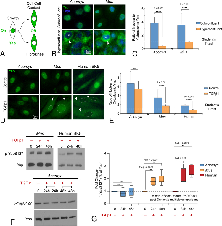
Figure 3: Acomys primary ear tissue fibroblasts evolved unique context-dependent regulation of Hippo pathway effector protein Yap.
(A) Diagram illustrating Yap localization as a readout for Hippo pathway activity.
(B, C) Immunofluorescent staining (B) and quantitation (C) for Yap (green) subcellular localization in Acomys and Mus primary ear tissue fibroblasts growing under homeostatic subconfluent and hyperconfluent cell densities in vitro (N ≥100 cells per treatment group). Asterisks indicate nucleus. Arrowheads indicate cytoplasm. Dapi serves as a nuclear localization marker. ns = not significant; data are mean ± SD; see also Figure S3B.
(D, E) Immunofluorescent staining (D) and quantitation (E) for subcellular localization of Yap after TGFβ1 (2ng/ml, 48h), arrowheads indicate cytoplasmic localization, dashed lines indicate nucleus: ns = not significant, N > 4 independent cultures; data are mean ± SD.
(F, G) Western blot (F) and quantitation (G) of pYap-S127 versus total Yap levels in during TGFβ1-mediated MF formation across species. Note that TGFβ1 stimulated Yap-S127 phosphorylation in Mus and human fibroblasts, but not in Acomys fibroblasts. ns = not significant. N ≥ 4 independent cultures; data are mean ± SD.

