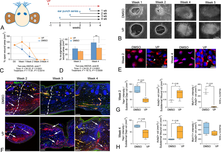
Figure 6: Yap activity is required for epimorphic ear regeneration and to prevent fibrosis in Acomys.
(A) Schematic diagram of chronic verteporfin (VP) treatment relative to ear punch timing in Acomys.
(B) Representative images of ear hole closure in verteporfin-treated (VP) and vehicle-treated (DMSO) Acomys. Dashed lines indicate border of open wound hole.
(C, D) Quantitation reveals significant attenuation of ear closure and decreased re-pigmentation in traumatic ear wounds due to VP (orange) relative to DMSO control (blue).
(E) VP treatment redistributes nuclear Yap to the cytoplasm in vivo at week 2 and maintained through week 4 (asterisks indicate nuclei, arrowheads indicate cytoplasmic Yap OFF), with attenuated in vivo Ctgf target labeling at week 1 and without body weight change (Figure S7F–J)
(F) Increased and persistent Acta2+/Myh11− MFs are present due to VP-treatment, and Acta2+/Myh11+ BV formation is delayed compared to control DMSO-treatment; note that microscopic views at week 3 indicate failure to close fully, with incomplete fusion and scab clearance (arrows, week 4).
(G, H) Quantitation for Acta2, Myh11 and Yap in VP-treated and DMSO-treated ear tissues at week 2 (G) and week 4 (H) traumatic ear repair: nuclear Yap is decreased, Acta2+/Myh11− MFs are increased, and Acta2+/Myh11+ vasculature formation is attenuated in VP-treated tissues: all data are mean ± SD.

