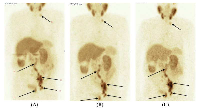
Figure 3.

(A–C) Male 87 year, adenoca G3; GlS = 8 (4 + 4). PSA 228.0 ng/mL. Evaluation of tumor extent. Three scans of WB-SPECT/CT acquisition after 1 h (A), 3 h (B) and 6 h (C) after radio-tracer i.v. administration. Pathological high uptake of [99mTc]Tc-PSMA-T4 is seen in primary PCa—left lobe of prostate (1), multiple lymph nodes: left obturator (2), left external and common iliac (3), paraaortic left and right (4) and the left neck (5). No bone lesions were detected in current PSMA, bone and CT scans.
