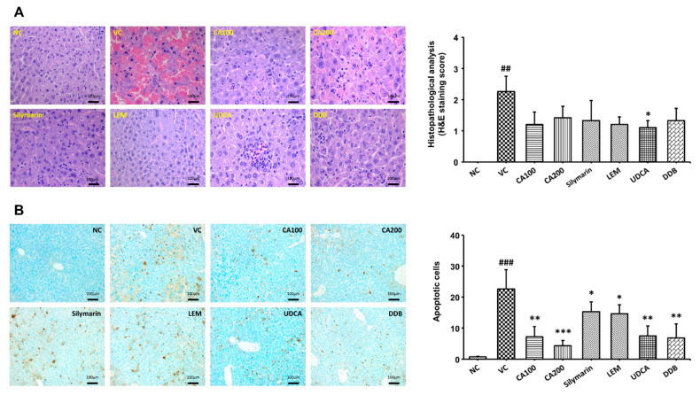
Figure 5.
Effect of CA-HE50 on histopathological changes in LPS/D-Gal-induced ALF animal model. Histopathological changes in the extracted liver tissue were confirmed by H&E and TUNEL analysis. Histopathological changes in liver tissue were quantified by lesion scores according to the severity of the lesion. (A) Representative images of each group in the histopathological lesion. Histopathological lesion scores are presented as a bar diagram. (B) Representative images of each group in apoptotic dead cells by TUNEL analysis. The numbers of apoptotic dead cells are presented as a bar diagram. NC, normal control; VC, vehicle control; CA100, 100 mg/kg CA-HE50; CA200, 200 mg/kg CA-HE50; Silymarin, 100 mg/kg silymarin; LEM, 200 mg/kg LEM; UDCA, 25 mg/kg UDCA; DDB, 200 mg/kg DDB. Data are represented as mean ± SD (## p < 0.01 or ### p < 0.001 vs. normal control; * p < 0.05, ** p < 0.01 or *** p < 0.001 vs. vehicle control).

