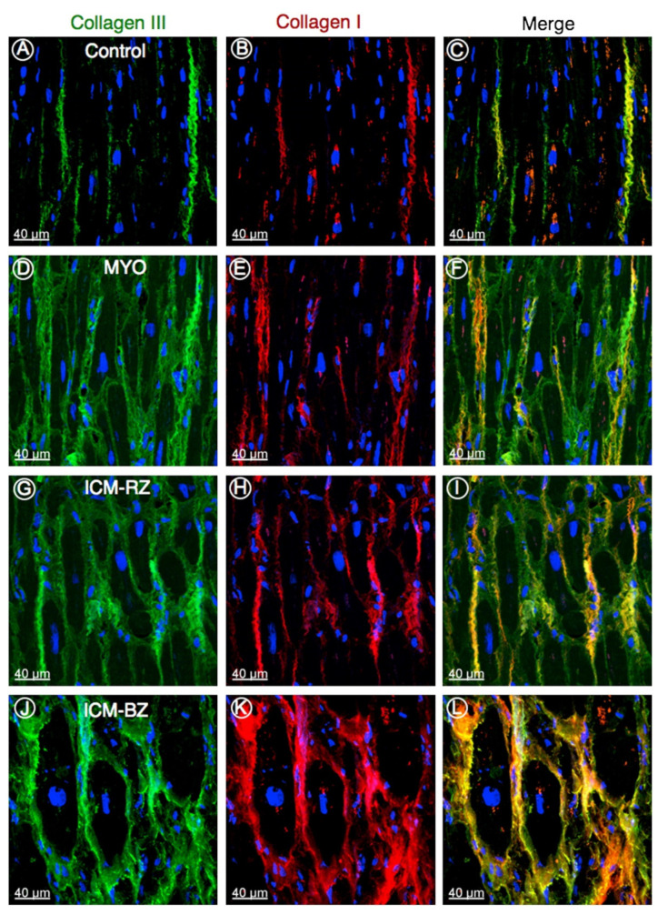
Figure 6.
Representative confocal images of collagen type III (green) and type I (red) in control (A–C), in explanted human heart tissue samples from patients with inflammatory cardiomyopathy (MYO; D–F), from patients with ischemic cardiomyopathy (ICM) from the remote zone (ICM-RZ; G–I) and border zone regions (ICM-BZ; J–L), demonstrating an increased accumulation of fibrillar collagens in diseased myocardium. Nuclei are stained blue with 4′,6-diamidino-2-phenylindole [105].

