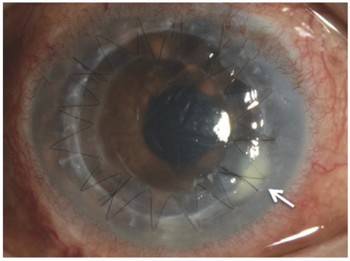
Figure 2.
Slit-lamp examination showing that small white infiltrates were observed at the border between the host and donor corneal graft. Adapted from Kitazawa K., Wakimasu K., Yoneda K., Iliakis B., Sotozono C., and Kinoshita S. A case of fungal keratitis and endophthalmitis post penetrating keratoplasty resulting from fungal contamination of the donor cornea. Am J Ophthalmol Case Rep. 2016; 5: 103–106 [32]. Figure 2A, Copyright (2016) with permission from Elsevier. License 5135721109911 on 25 August 2021.

