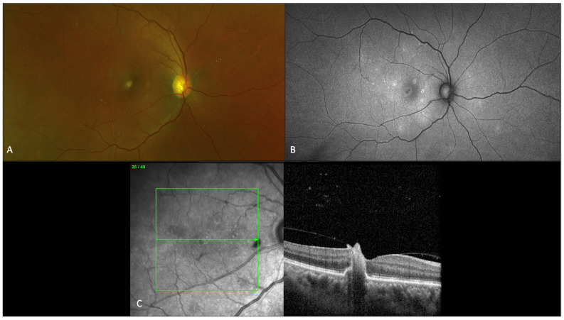
Figure 4.
Imaging findings for a 56-year-old patient with fungal chorioretinitis, showing characteristic fundus, autofluorescence, and optical coherence tomography (OCT) macula findings. (A) Fundus photograph of right eye showing multiple small chorioretinal white lesions predominantly in the macula. (B) Fundus autofluorescence showing large area of hypoautofluoroescence in the fovea and multiple areas of scattered punctate hyperautofluoresence lesions in areas of early retinal pigment epithelium loss. (C) OCT macula findings over the foveal chorioretinal lesion depicting infiltration from choroid through retinal layers into the vitreous with focal areas of traction.

