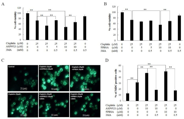
Figure 8.
Verification of autophagy as a major pathway of AKR1C1- and 1C3-regulated cell death. (A) KATO/DDP cells were treated together with cisplatin and ASP9521 with or without 3MA at 0.5 mM. Cell viability was assessed by using trypan blue cell exclusion assay. (B) KATO/DDP cells were treated together with cisplatin and 5PBSA with or without 3MA at 0.5 mM, and cell viability was assessed by using trypan blue cell exclusion assay. (C) KATO/DDP cells were treated together with cisplatin and ASP9521 or 5PBSA at 10 µM with or without 3MA at 0.5 mM. Autophagic vacuoles were stained with MDC dye and visualized under fluorescent microscope. (D) Graphical representation of % of MDC-positive cells in KATO/DDP. Data are presented as mean ± SD; ** represents p < 0.01.

