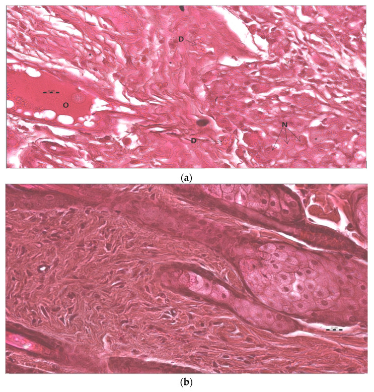
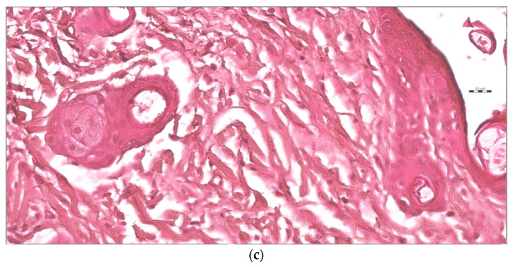
Figure 13.
(a) This is micro photo of negative control show severe tissue damage in the form of necrosis due to the absence of nuclei (N), degeneration in form of vacuoles (D), as well as occlusion of blood vessels by large spots of hyaline material (O). Stain is H&E, magnification is 400×, and scale bar is 20 µm. (b). The standard sample shows an almost normal tissue sample. Stain is H&E, magnification is 400×, and the scale bar is 20 µm. (c). Test tissue samples treated with AgNPs loaded chitosan-based gels show almost normal tissue sample. Stain is H&E, magnification is 400×, and scale bar is 20 µm.


