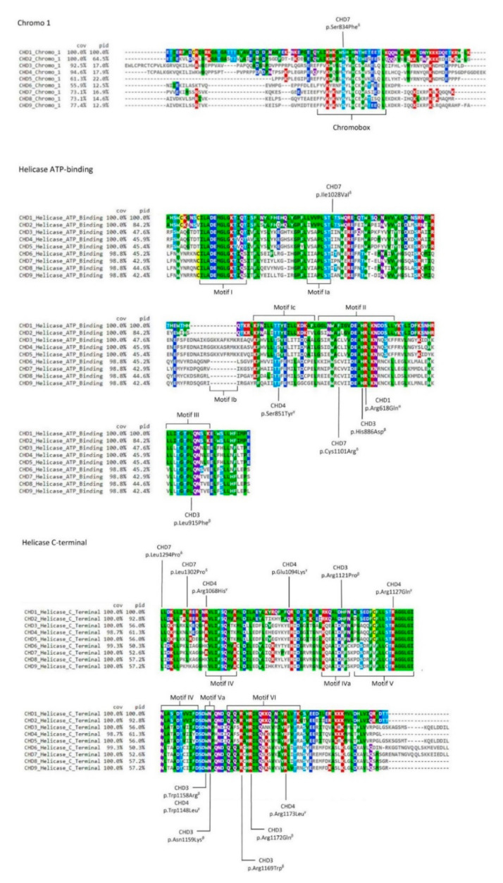
Figure 3.
Scheme of CHD paralogues with pathogenic variants obtained from the ClinVar database (https://www.ncbi.nlm.nih.gov/clinvar. Accessed on 2 November 2020) [63] also described in the literature (Table 1) for domains Chromo 1, Helicase ATP-binding, and Helicase C-terminal. Letters α, β, γ, and δ represent the clinical phenotypes associated with each pathogenic variant (see Table 1). Chromobox and motifs I, Ia, Ib, Ic, II, III, IV, IVa, V, Va, and VI are displayed. The colour scheme represents the variable amino acid positions after using the multiple alignment viewer MView (COV—% coverage; ID—% identity). The mapping of the chromobox and the motifs was performed according to references [27,28,51,85,86].

