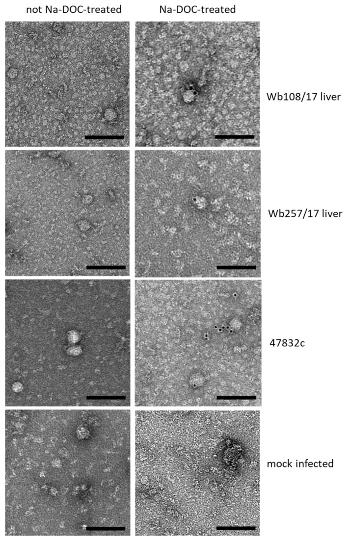
Figure 4.
Analysis of particle morphology by immunogold staining and transmission electron microscopy. Samples, as indicated on the right side of the images per row, were treated (right image) with sodium deoxycholate (Na-DOC) to remove the quasi-envelop from HEV, or not treated (left image). Immunogold-staining was done using an anti-HEV capsid protein-specific monoclonal antibody together with a gold-labeled secondary antibody. Black dots represent the gold particles. Negative staining with uranyl acetate. Scale bar: 100 nm.

