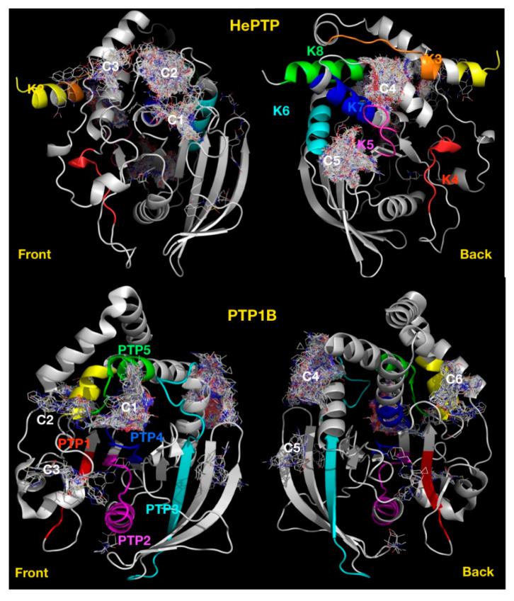
Figure 6.
Blind docking cluster comparison between HePTP and PTP1B. The structure of HePTP (top) is shown as a grey cartoon, key functional KIM specific motifs [33] have been coloured on the HePTP structure, K2 in yellow, K3 in orange, K4 in red, K5 in magenta, K6 in cyan, K7 in blue, K8 in green. Compounds are shown as lines. Ligand clusters with more than ten compounds are numbered: C1, active site; C2, open form pocket; C3, substrate-binding site; C4, centred on K3, K5 and K7 motifs; C5, centred on K5–K7 motifs. The structure of PTP1B is shown as a grey cartoon (bottom), front view and back view. Clusters are numbered: C1, active site; C2, secondary pTyr site; C3, centred on L41; C4, allosteric binding site; C5, centred on V155; and C6, centred on E252. Key PTP motifs [33] have been coloured on the structure: PTP1 in red; PTP 2, magenta; PTP 3, cyan; PTP 4, blue; PTP 5, green; and PTP6, yellow.

