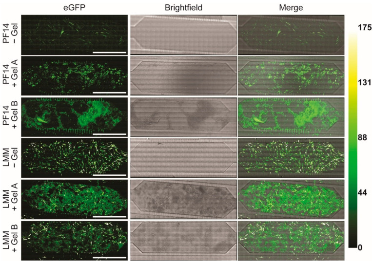
Figure 3.
Expression of eGFP mRNA in the absence and presence of colloidal gelatin. Confocal microscopy images of eGFP expression in MC3T3 cells 24 h post-transfection. The conditions without gelatin were used to assess the impact of colloidal gelatin on transfection efficiency. The Green-Hot LUT on the right depicts the eGFP intensity. Brightness and contrast were individually adjusted for all images. Scale bars represent 1000 µm. eGFP, enhanced green fluorescent protein.

