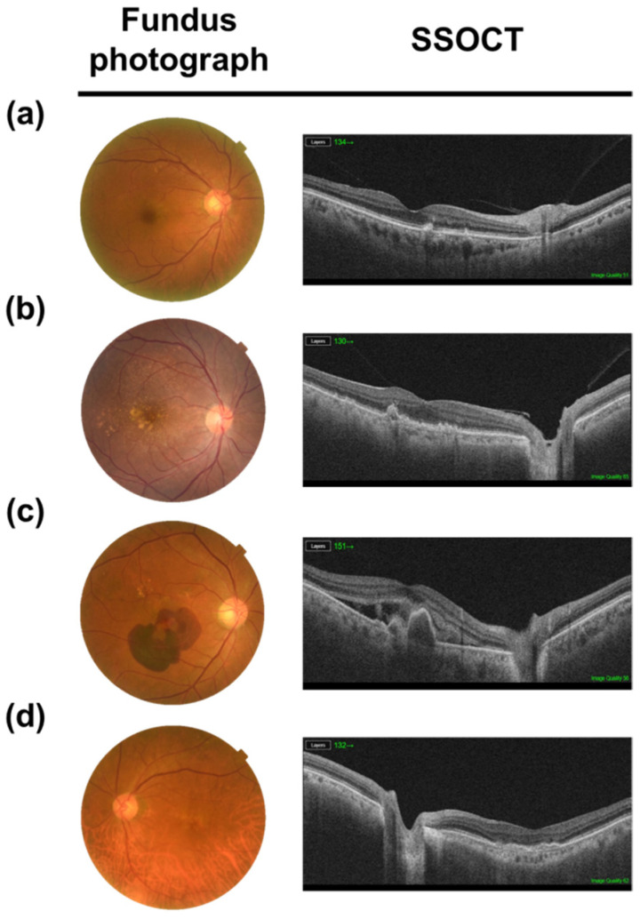
Figure 1.
Multimodal image of age-related macular degeneration (AMD). Color fundus photograph and swept source optical coherence tomography (SS-OCT) images show the feature of early, intermediated AMD, neovascular AMD and geographic atrophy (a,b). Non-neovascular AMD (dry AMD): small and intermediate soft drusen. (c) Neovacular AMD (wet AMD): submacular hemorrhage, subretinal fluid and pigment epithelial detachement. (d) Geographic atrophy: retinal pigment epithelial pigment and photoreceptor atrophy at fovea.

