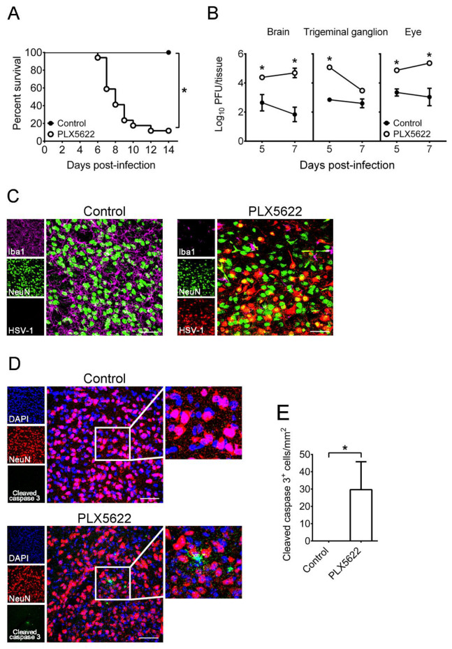
Figure 2.
The effects of PLX5622 on HSV-1 lethality, tissue viral loads, and brain infected neurons as well as neuron apoptosis of mice. (A) Mice were fed with control chow (n = 16) or the chow containing PLX5622 (n = 17), infected with HSV-1, and monitored for survival. (B) The indicated organs and tissues of control and PLX5622-treated mice were harvested on the indicated days for virus titration and on day 7 post-infection for brain sections (C,D) before immunofluorescence staining with antibodies against Iba1, NeuN, HSV-1, or cleaved caspase 3. Scale bar, 50 μm. In panel C, right panels are the merged and magnified images of three left panels. In panel (D), middle panels are the merged and magnified images of three left panels, and right panels are the magnified images of indicated areas show in middle panels. (E) The number of leaved caspase 3+ cells in images (0.3 × 0.3 mm) were counted. Results shown in panels (C–E) are the temporal region and the representative of at least 3 samples per group from two independent experiments. The data represent means ± SEM (error bars) of 3–6 samples per data point obtained from at least two independent experiments in panels (B,E). *, p < 0.05, compared between the indicated groups (A,E) or with the control group on the same day (B).

