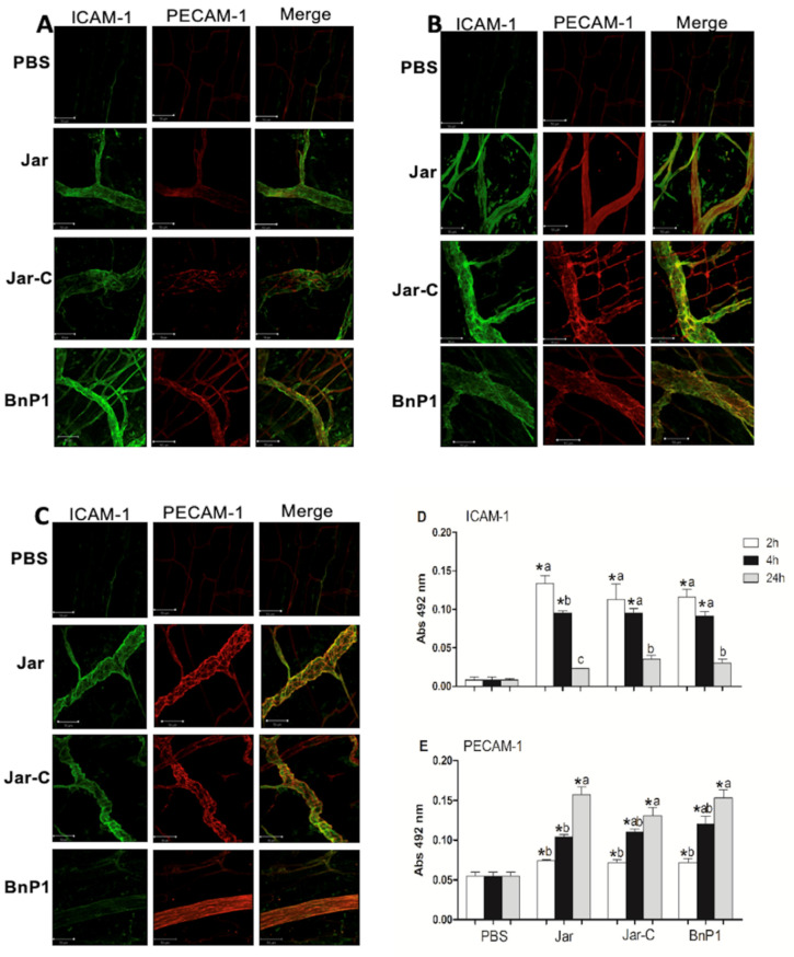
Figure 1.
Expression of ICAM-1 and PECAM-1 in the cremaster muscle at different time points. The panels present photomicrographs of immunofluorescence labeling analyzed using confocal microscopy at 2 h (A), 4 h (B) and 24 h (C) after the subcutaneous injection of PBS, Jar, Jar-C or BnP1 (0.5 μg/100 μL) in the mouse scrotum. Bar, 50 µm. Quantification of the protein levels of ICAM-1 (D) or PECAM-1 (E) in homogenates of cremaster muscle determined using ELISA. Absorbance was measured at 492 nm. The results are presented as the means ± S.E.M. (n = 5). * p < 0.05 compared to the PBS group at any time point. Different letters (a, b, c) in the same group represent significant differences (* p < 0.05).

