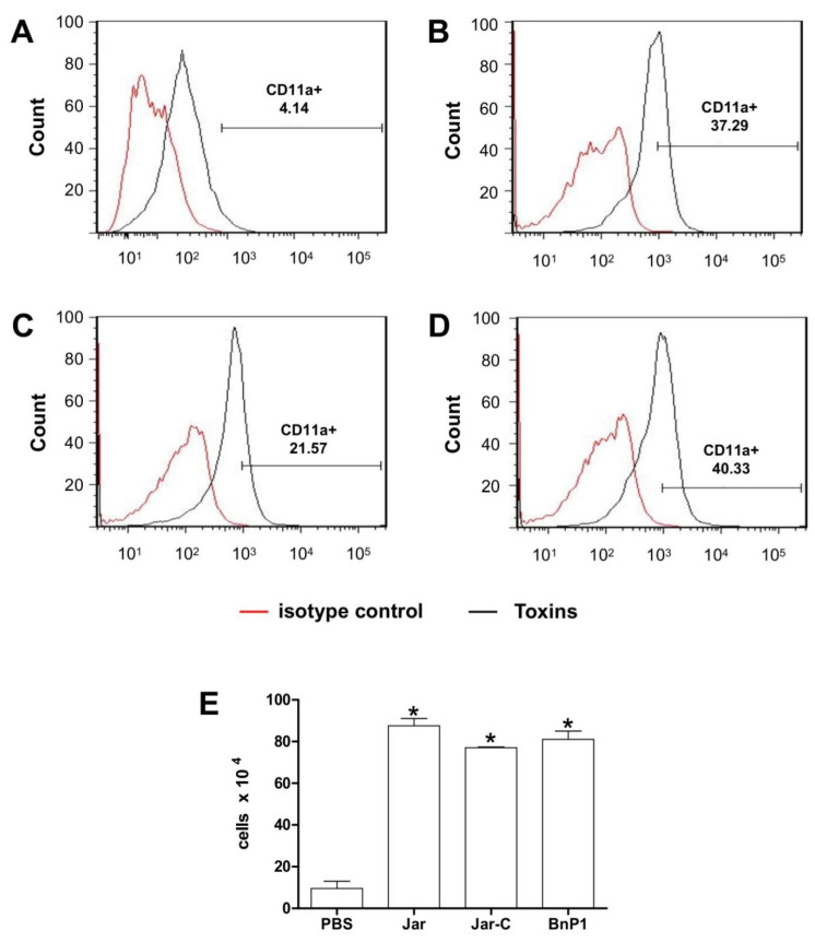
Figure 3.
Expression of CD11a on the surface of leukocytes present in the peritoneal exudate of mice injected with different toxins. Peritoneal exudate cell suspensions were obtained within 4 h after the injection of PBS (A), Jar (B), Jar-C (C) or BnP1 (D) (2 μg/300 μL). Cells were incubated with anti-CD11a-FITC antibodies or isotype control-FITC. All incubations with anti-CD11a-FITC were performed on duplicate samples and analyzed using flow cytometry. Histograms are representative of one experiment. The bar graph shows the average numbers of CD11a-positive cells in each experimental group ± SD from three independent experiments (E). * p < 0.05 compared to the PBS group.

