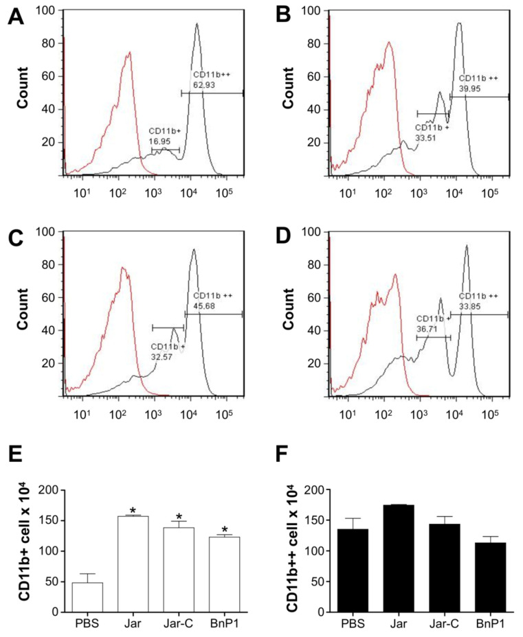
Figure 4.
Expression of CD11b on the surface of leukocytes present in the peritoneal exudate of mice injected with different toxins. Peritoneal exudate cell suspensions were obtained within 4 h after the injection of PBS (A), Jar (B), Jar-C (C) or BnP1 (D) (2 μg/300 μL). Cells were incubated with anti-CD11b-FITC antibodies or isotype control-FITC. All incubations with anti-CD11b-FITC were performed on duplicate samples and analyzed using flow cytometry. Histograms are representative of one experiment. The bar graph shows the average numbers of positively stained cells designated as CD11b+ (E) or CD11b++ (F) in each experimental group ± SD from three independent experiments. * p < 0.05 compared to the PBS group.

