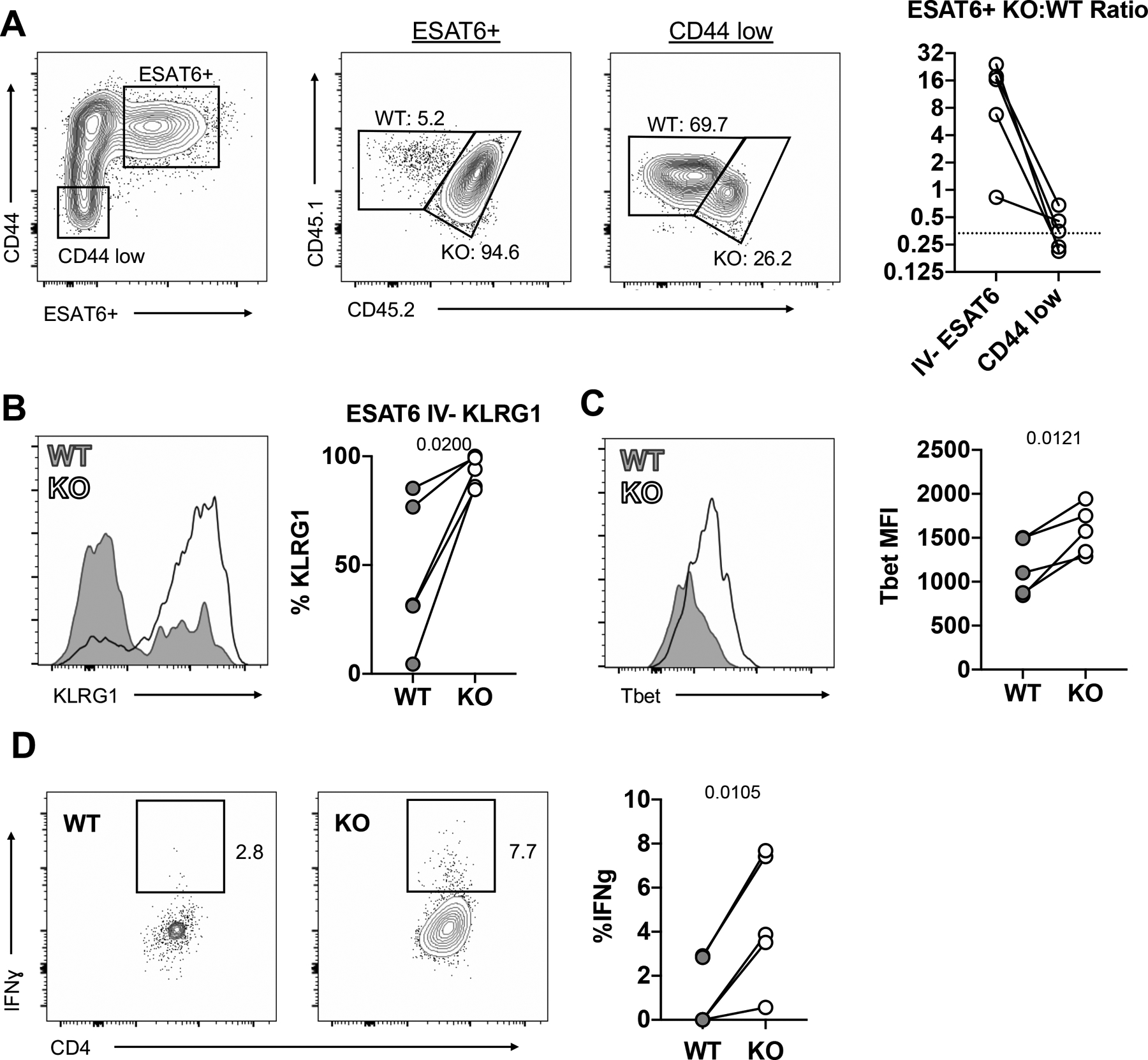
Figure 4: TGFβ acts intrinsically on CD4 T cells to regulate Th1 cell differentiation and cellularity.

Mixed WT:TGFβR.KO bone marrow chimeric mice infected with 50–100 CFU of Mtb. (n=5) A) TGFβR.KO:WT ratio of ESAT-6-specific cells and CD44 low naïve cells. B) Percentage of parenchymal ESAT-6 specific CD4 T cells that are KLRG1+ in WT vs TGFβR.KO mice C) T-bet MFI of parenchymal ESAT-6 specific CD4 T cells. D) Ex-vivo IFNγ production by parenchymal ESAT-6-specific cells. Lines connect WT and KO cells within the same chimeric animals. Single-group comparisons by paired t test. Data are representative of three independent experiments. Points are individual mice, lines connect WT and KO cells within the same chimeric animals., Data are presented as mean ± SD. *p < 0.05, **p < 0.01, ***p < 0.001, ****p < 0.0001. See also Figure S4 and S5.
