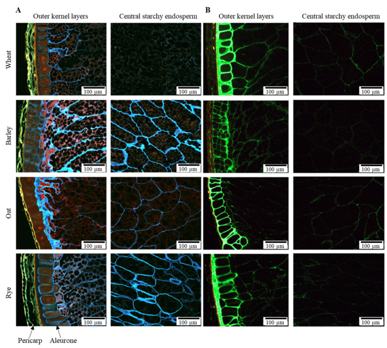
Figure 2.
Epifluorescence microscopy pictures of outer kernel layers and central starchy endosperm of wheat, barley, oat and rye. (A) β-glucan is stained with Calcofluor (blue), protein with Acid Fuchsin (red). The pericarp is visible by autofluorescence (yellow). (B) Arabinoxylan is stained with an inactive fluorescently labelled xylanase (green) [35]. Adapted from Dornez et al. (2011) [35], reprinted with permission from Elsevier.

