FIG. 1-4.
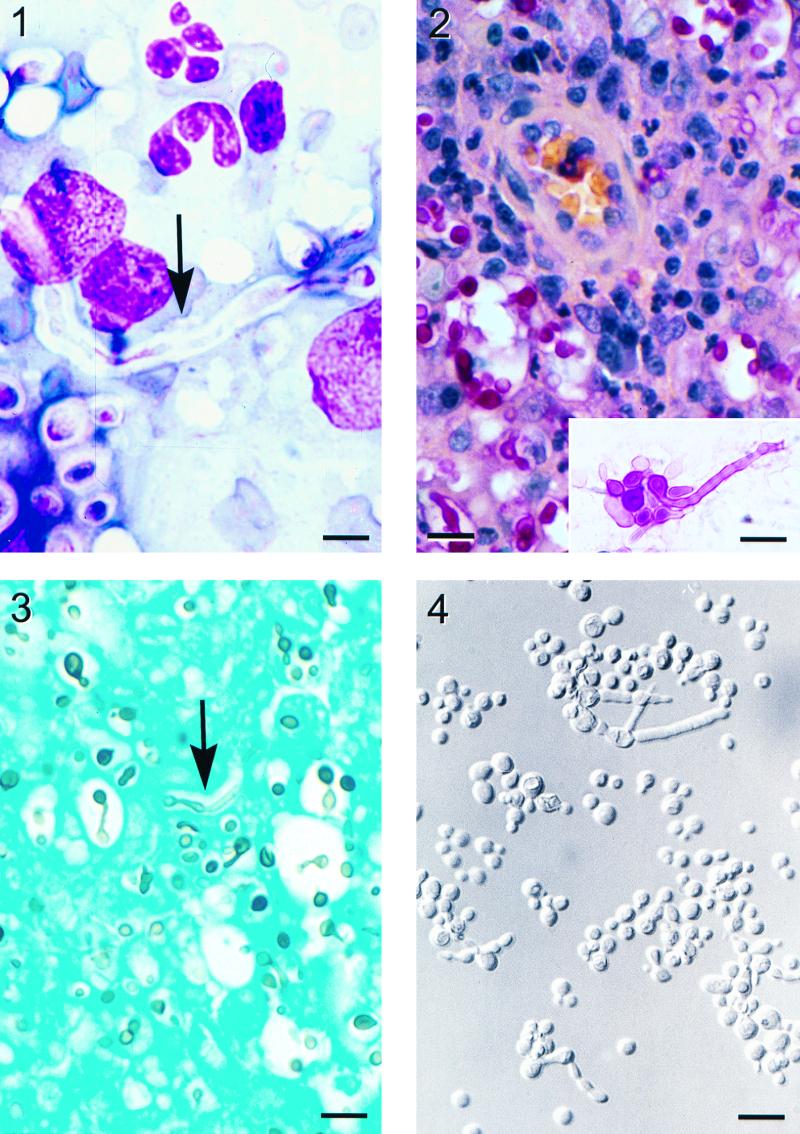
FIG. 1. Wright stain of a nasal tissue impression smear demonstrating chronic inflammatory cells, encapsulated yeast cells, and a long hyphal element (arrow) extending from a yeast cell. Bar = 8 μm. FIG. 2. Nasal granuloma tissue. A capillary with perivascular cuffing of lymphocytes and plasma cells is seen among many histiocytes and yeast cells that are positively stained with mucicarmine. Bar = 30 μm. (Inset) Some yeast cells have germ tube or hyphal elements extending from them. Bar = 20 μm. FIG. 3. Gomori's methemine silver stain of nasal granuloma tissue. Hyphae with parallel walls (arrow) are occasionally seen among the yeast cells. Bar = 27 μm. FIG. 4. Yeast cells grown on YEPD agar at 35°C for 5 days showing budding yeast, yeast cells in short chains, and yeast cells with hyphal elements. Bar = 13 μm. All photo images were digitized with a CoolScan II scanner (Nikon, Inc., Tokyo, Japan) and formatted with Adobe Photoshop 5.0 software (Adobe Systems Incorporated, San Jose, Calif.).

