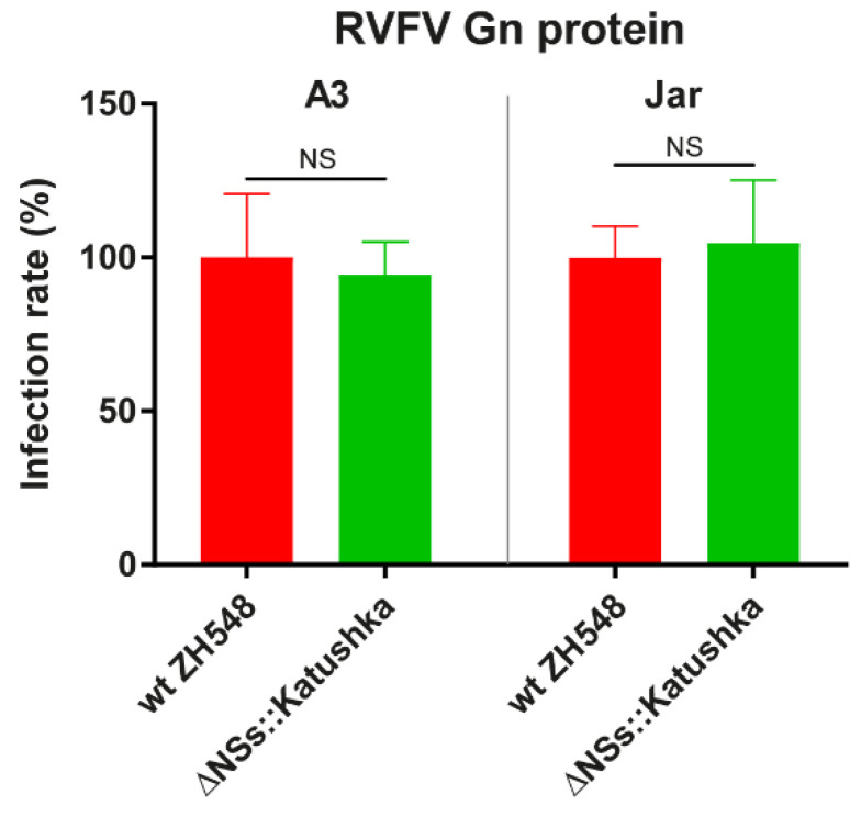
Figure 2.
Rift Valley fever virus (RVFV) Gn protein expression in the A3 and Jar cell lines. The cell lines were infected at a multiplicity of infection of 1 and the Gn protein was detected by using an anti-RVFV Gn monoclonal antibody, and a secondary anti-mouse antibody conjugated to Alexa Fluor 488. Cytation 5 Cell Imaging Multi-Mode Reader identified green fluorescent protein (GFP)- or 4′,6-diamidino-2-phenylindole (DAPI)-expressing cells and quantified the fluorescence intensity in each well. Experiments were performed in triplicate and repeated twice. The bar plus error bar indicates the mean ± standard deviation. Statistical significance was determined by multiple t-test. p values are indicated (NS = not significant).

