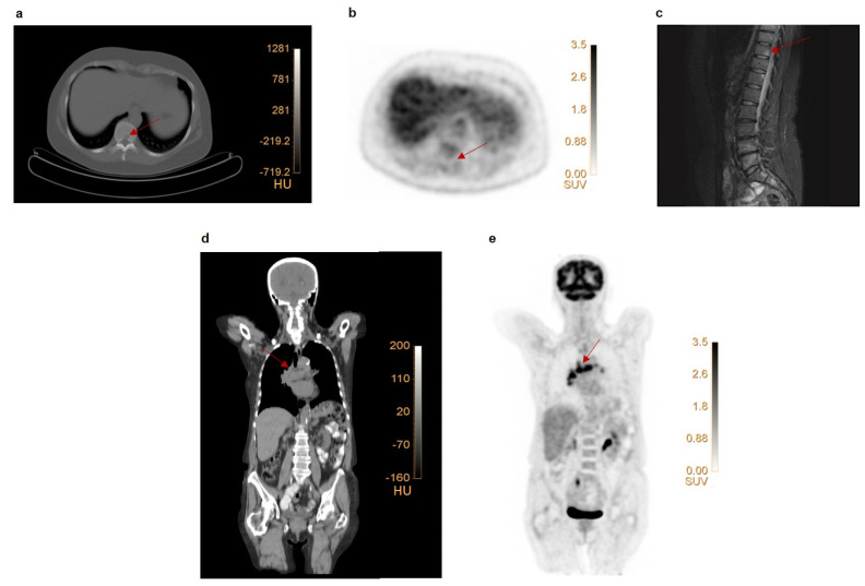
Figure 1.
Examples of false negative and false positive lesions on FDG PET. (a–c) Patient with primary ER+ breast cancer with faint uptake in the primary tumor (SUVmax 2.3). Low-dose CT (a) revealed a lytic lesion in the 10th thoracic vertebra (Th10) without enhanced FDG uptake (b). An MRI scan (c) revealed multiple vertebral metastases (Th4, Th11, Th12, L4, L5), including the one at Th10. This lesion was classified as false negative on FDG PET. (d–e) Patient with multiple mediastinal FDG avid, suspect lymph nodes. Coronal section of a low-dose CT-scan (d) and FDG PET scan (e). Endobronchial ultrasound–guided biopsy of 3 mediastinal lymph nodes showed reactive cells. These lesions were, therefore, classified as false positive on FDG PET.

