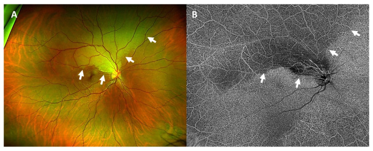
Figure 1.
Wide-angle fundus photography of the right eye (A) showed a pale, edematous area corresponding to the area of arterial occlusion (white arrows). In optical coherence tomography angiography (B) the area of arterial occlusion was seen as a hypoautofluorescent area with a missing blood supply (white arrows).

