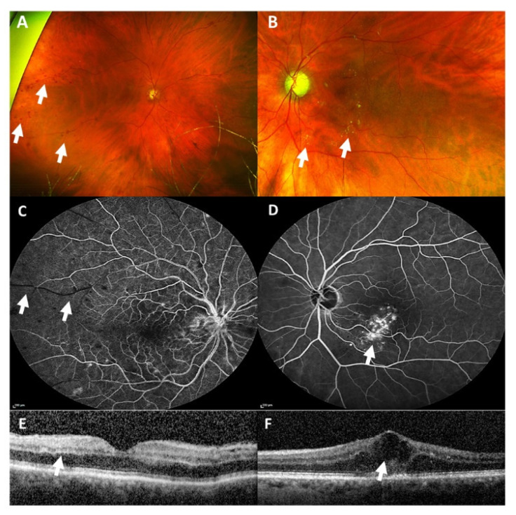
Figure 2.
Wide-angle fundus photography (A,B) revealed multiple intraretinal hemorrhages in the right eye (white arrows, A) and hard exudates, seen as yellowish circumscribed spots, in the left eye (white arrows, B). Fluorescein angiography (C,D) showed an arterial capillary non-perfusion (white arrows, C) in the right eye and vessel leakage inferior of the macula in the left eye, seen as hyperfluorescent non-well circumscribed area (white arrow, D). Optical coherence tomography (E,F) revealed a hyperreflectivity of inner retinal layers as a sign of an arterial occlusion (white arrow, E) in the right eye, and a cystoid macular edema (white arrow, F) in the left eye.

