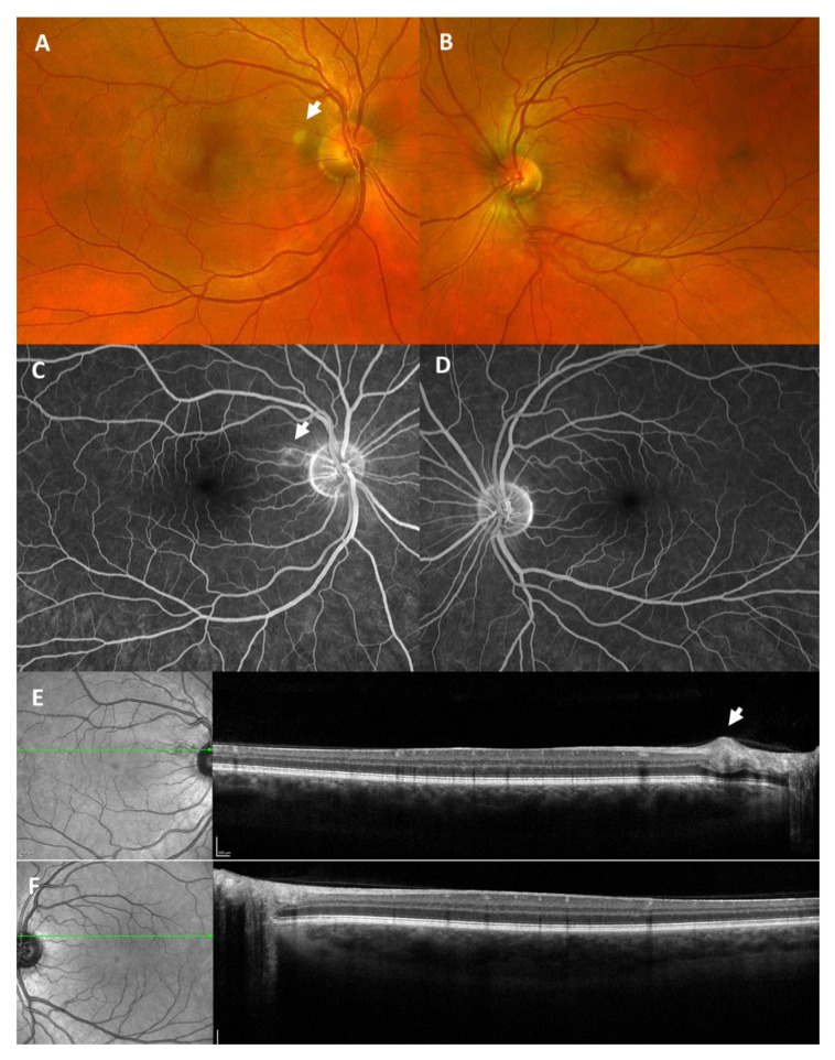
Figure 5.
Fundus photography (A,B) revealed a cotton-wool spot temporal of the optic disc (white arrow) in the right eye (A) and was normal in the left eye (B). Fluorescein angiography (C,D) showed a hyperfluorescent spot (white arrow) corresponding to the cotton-wool spot in the right eye (C) and was normal in the left eye (D). Optical coherence tomography (E,F) revealed a circumscribed swelling of the retinal nerve fiber layer (white arrow) corresponding to the cotton-wool spot in the right eye (E) and was normal in the left eye (F). The green arrow indicates the position the OCT image was taken.

