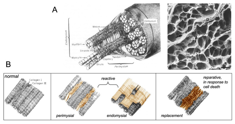
Figure 1.
Fibrous skeleton of the heart and patterns of fibrosis. (A) Schematic illustration of the normal collagen matrix, illustrating perimysial fibrous tissue enveloping myocyte bundles and endomysial fibrous tissue in between strands of myocytes within bundles (left panel). Acellular preparation after digestion of cells showing perimysial sheets (p) and endomysial septae (e). (B) Schematic representation of the normal ECM (left panel), endomysial and perimysial fibrosis (middle panel), also called reactive fibrosis, and replacement/ reparative fibrosis secondary to myocyte death (right panel), adapted from [1].

