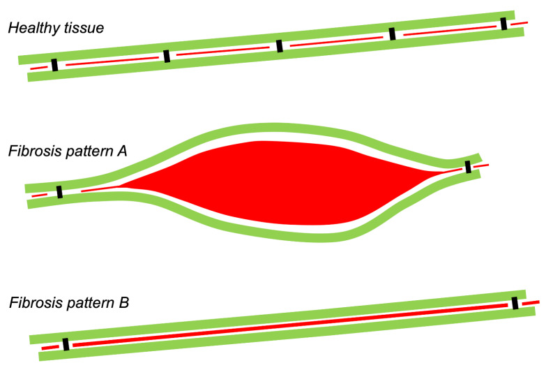
Figure 2.
Relation between amount of fibrous tissue and electrical consequences. Conceptual diagram of two strands of longitudinally coupled myocytes (green) connected by sparse, discrete transverse connections (black). In between transverse connections, myocytes are separated by (endomysial) fibrous tissue (red). In fibrosis pattern A, a large area of fibrous tissue separates the strands, and in pattern B, the thickened endomysial septum. Both patterns have reduced transverse connectivity by the same degree, and will therefore have similar consequences for propagation, although pattern B is detected more readily in a histological analysis.

