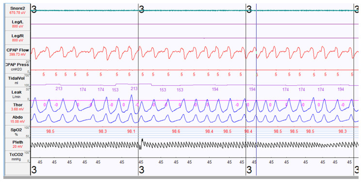
Figure 3.
Polysomnography tracing during PAP titration study. Each tracing noted from top to bottom are as follows: snoring microphone, electromyogram of the bilateral tibia on the left and then the right leg, CPAP flow showing air flow, CPAP pressure setting of 5cmH20, tidal volume, air leak in L/min (currently at 0 L/min), chest and abdominal wall motion (respiratory inductive plethysmography), arterial oxygen saturation value and waveform, and transcutaneous carbon dioxide waveform (shown at 45 mmHg).

