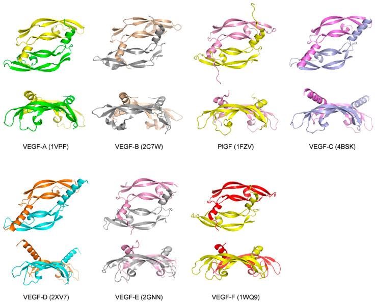
Figure 1.
Structures of the receptor-binding domains of VEGF family members. The top representation shows the view along the two-fold symetry axis of VEGF, while the bottom representation shows a perpendicular view. PDB codes are given in parenthesis. For VEGF-C (4BSK), only the growth factor is shown, while the structure includes a receptor fragment.

