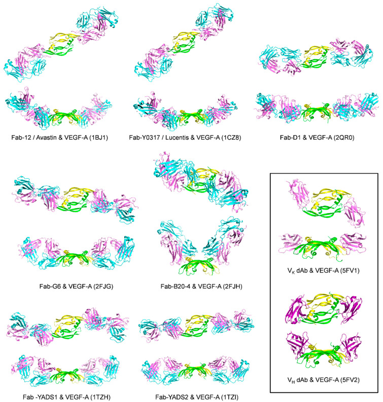
Figure 7.
Ribbon representation of antibody/VEGF-A complexes. VEGF-A is colored in green and yellow, and the antibodies are colored in various shades of pink and blue. The top representation shows the front view for each complex, while the bottom representation shows the side view. The insert shows the domain antibody (dAb)/VEGF-A complexes. PDB codes are given in parenthesis.

