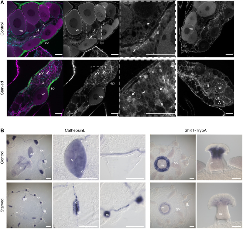
Fig. 7. Perturbations to gastroderm cell types in response to starvation.
(A) Confocal sections through gonads from control and starved medusae with cell morphology revealed by phalloidin staining of cell boundaries (magenta/gray). The first panel of each row shows costaining of nuclei with Hoechst (blue) and endogenous GFP4 (green) in the outer epidermis (epi). Vitellogenic oocytes (vo) are largely absent during starvation, leaving mostly previtellogenic oocytes (po). The gastroderm (g) is heavily reorganized, with evidence of active phagocytosis (vesicles arrowed) and disintegrating oocytes (asterisks). The third panel in each row is a higher magnification of the boxed area in the second panel, and the fourth panel shows a second example gonad for each condition. Scale bars, 50 μm. (B) In situ hybridization visualization of example cell-types (left) and genes (right) affected by starvation. The general GD cell marker CathepsinL confirms extensive reorganization of the digestive gastroderm in starved animals, especially its collapse into central regions of the strongly reduced, oocyte-depleted, gonad, and fragmentation in the circular canal; expression of the gland cell A/B marker “ShKT and trypsin domain protein A” (ShKT-TrypA) in the manubrium consistent with its strong down-regulated following starvation. Gonads, go; manubrium, m; tentacle bulbs, tb; gastroderm, g; circular canal, cc. Scale bars, 200 μm.

