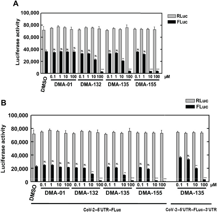
Fig. 6. Vero E6 cells were cotransfected with luciferase constructs and cultured with various concentration of small molecule.
(A) Vero E6 cells were cotransfected with CoV-2-5’UTR-FLuc-3’UTR. (B) Vero E6 cells were cotransfected with Cov-2–5′UTR–FLuc (left), and no change in inhibition was observed when compared to the presence of 3′-UTR (right, DMA-135). Luciferase activity was measured 2 days later. Mean values and SDs from three independent experiments are shown in the bar graphs. ***P < 0.001; **P < 0.01; N., not significant relative to the DMSO control, except for the 5′UTR-FLuc-3′UTR comparison in (B), which is relative to DMA-135 at 0.1 μM.

