. 2021 Nov 8;10:e67952. doi: 10.7554/eLife.67952

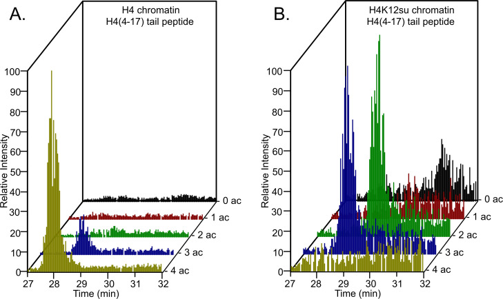
Figure 3. Comparison of H4 tail acetylation by p300 in chromatinized plasmid templates with activator Gal4-VP16.

(A) Extracted ion chromatograms of all H4(4–17) tryptic peptides obtained after SDS-PAGE resolution and in-gel trypsination of acetylated chromatin containing wild-type (wt) H4. (B) Extracted ion chromatograms of all H4(4–17) tryptic peptides obtained after SDS-PAGE resolution, in-gel desumoylation and trypsination of acetylated chromatin containing H4K12su. The extracted m/z of each spectrum is centered on the [M + 2 H]2+ precursor ion.

Figure 3—source data 1. Excel file listing all mass spectral data plotted in Figure 3, Tables 14; and Figure 3—figure supplements 27.
elife-67952-fig3-data1.xlsx (478.6KB, xlsx)

Figure 3.

Figure 3—figure supplement 1. Coomassie-stained SDS-PAGE of histone acetylation assay on chromatinized plasmids containing wild-type (wt) H4 or H4K12su with p300 and activator Gal4-VP16.
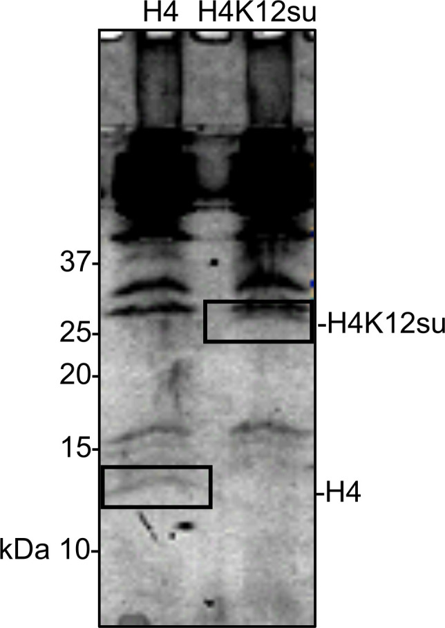
Figure 3—figure supplement 1.

Gel bands excised for tandem mass spectrometry (MS-MS) analysis are indicated.
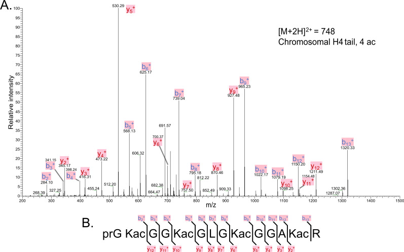
Figure 3—figure supplement 2. Tandem MS of tetra-acetylated tryptic peptide H4(4–17).

Figure 3—figure supplement 2.

(A) Representative tandem mass spectrometry (MS-MS) spectrum of tetra-acetylated tryptic peptide H4(4–17) generated after in vitro acetylation of chromatinized plasmids containing wild-type (wt) H4 with p300 and activator Gal4-VP16. (B) Peptide fragment-ion map of the tetra-acetylated H4(4–17) peptide indicating all ions identified over three spectra.
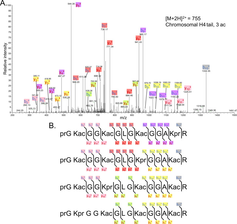
Figure 3—figure supplement 3. Tandem MS of tri-acetylated tryptic peptide H4(4–17).

Figure 3—figure supplement 3.

(A) Representative tandem mass spectrometry (MS-MS) spectrum of tri-acetylated tryptic peptide H4(4–17) generated after in vitro acetylation of chromatinized plasmids containing wild-type (wt) H4 with p300 and activator Gal4-VP16. (B) Peptide fragment-ion maps of the four possible tri-acetylated H4(4–17) peptide patterns, indicating all ions identified over three spectra.
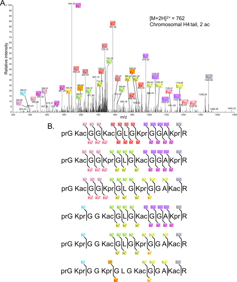
Figure 3—figure supplement 4. Tandem MS of di-acetylated tryptic peptide H4(4–17).

Figure 3—figure supplement 4.

(A) Representative tandem mass spectrometry (MS-MS) spectrum of di-acetylated tryptic peptide H4(4–17) generated after in vitro acetylation of chromatinized plasmids containing wild-type (wt) H4 with p300 and activator Gal4-VP16. (B) Peptide fragment-ion maps of the six possible di-acetylated H4(4–17) peptide patterns, indicating all ions identified over two spectra.
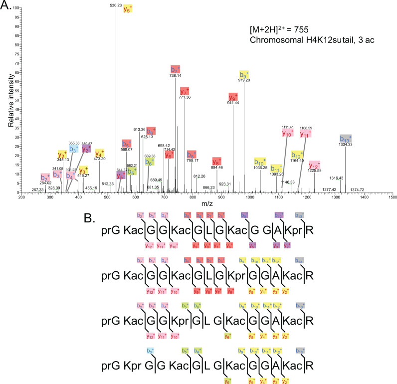
Figure 3—figure supplement 5. Representative tandem mass spectrometry (MS-MS) spectrum of tri-acetylated tryptic peptide H4(4–17) generated after in vitro acetylation of chromatinized plasmids containing H4K12su with p300 and activator Gal4-VP16 followed by in-gel desumoylation.

Figure 3—figure supplement 5.

(B) Peptide fragment-ion maps of the four possible tri-acetylated H4(4–17) peptide patterns, indicating all ions identified over three spectra. Acetylation on K12 in H4K12su is not possible due to presence of SUMO-3 at K12.
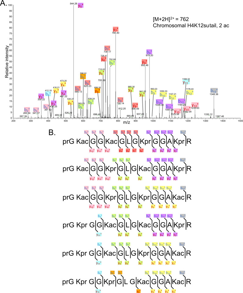
Figure 3—figure supplement 6. Representative tandem mass spectrometry (MS-MS) spectrum of di-acetylated tryptic peptide H4(4–17) generated after in vitro acetylation of chromatinized plasmids containing H4K12su with p300 and activator Gal4-VP16 followed by desumoylation.

Figure 3—figure supplement 6.

(B) Peptide fragment-ion maps of the six possible di-acetylated H4(4–17) peptide species, indicating all ions identified over three spectra.
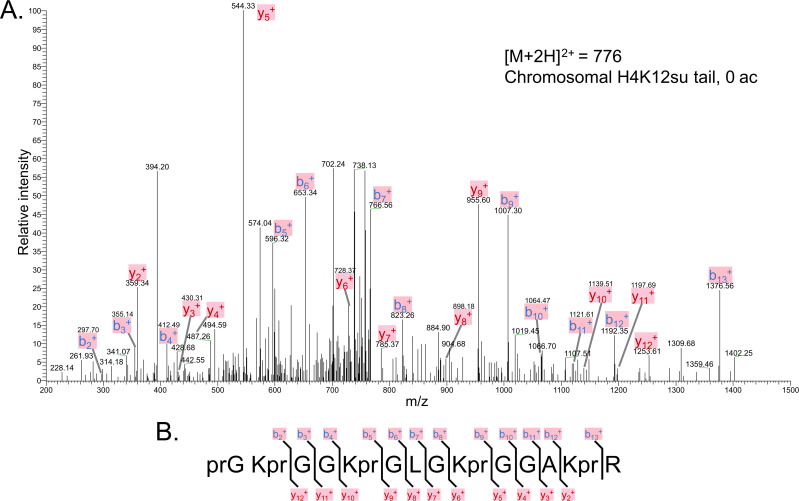
Figure 3—figure supplement 7. The tandem mass spectrometry (MS-MS) spectrum of unacetylated tryptic peptide H4(4–17) after in vitro acetylation of chromatinized plasmids containing H4K12su with p300 and activator Gal4-VP16 followed by desumoylation.

Figure 3—figure supplement 7.

(B) Peptide fragment-ion map of the unmodified H4(4–17) peptide, indicating identified ions.