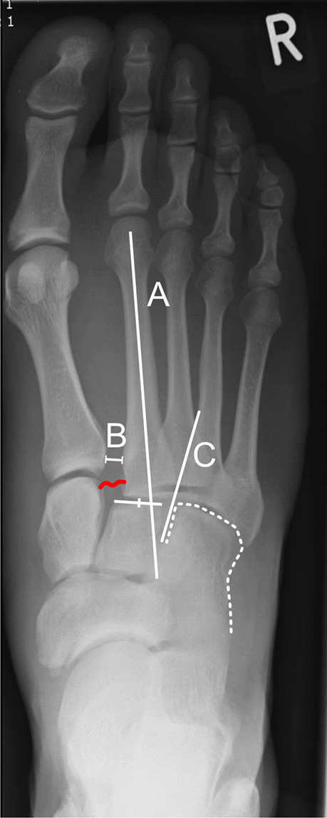
Fig. 1.

Radiologic criteria indicating if a Lisfranc injury is present in a plain dorsoplantar radiography, as published by Buehren [5]. Buehren A: The shaft axis of the second metatarsal bone physiologically points at the center of the second cuneiform. In this example, the axis does not project at the center, suggesting a Lisfranc injury. Buehren B: The distance of the basis of the first and second metatarsal bone should not exceed 3 mm. In this example, the distance was 7.5 mm. Buehren C: The tangent of the medial basis of the fourth metatarsal bone should exactly be in line with the medial cortex of the cuboid, as seen in this example. The red curved line indicates the position of the Lisfranc ligament between C1 and M2, which is suspected to be torn in this example
